A New Technique to Scan the Human Body
Engineers at Lancaster University and scientists at the Institute of Food Research are developing a quick, safe and non-invasive scanner to measure the composition of the human body.
The prototype scanner uses two imaging techniques to predict the amount of fat present by estimating both the volume and water content of the body. It measures the outer shape of the body and could estimate regional variations in composition such as fat distribution.
“Body composition is an indicator of an individual’s nutritional status. It can be used to monitor child development, pregnancy, and changes during diet and exercise regimes. In addition to clinical applications, our system could also become a feature of leisure centres, allowing clients to see how their shape and composition changes through exercise”, says Dr Henri Tapp, from the Institute of Food Research.
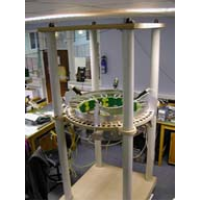
The specially designed scanning cubicle is fitted with digital cameras and light projectors to map the surface contours of the body, to give body volume. An array of coils is used to map the internal electrical conductivity, to give water content. The camera and coils are fitted to a sliding sensor ring, designed to scan the whole body as a series of horizontal ‘slices’.
Although the initial aim is to predict percentage fat content, the system could potentially also provide regional information. This includes estimates for individual body segments, or differentiating between surface and internal fat within the torso. The surface data also provides anthropometric measures of body shape, such as android (‘apple-shaped’) and gynoid (‘pear-shaped’) fat distributions. This could be clinically useful, for example in epidemiological studies comparing and relating body shapes to health risks such as heart disease.
Traditional measurement methods are time-consuming and inconvenient. For example, calculating body volume involves repeatedly submerging an individual in water, while simultaneously estimating lung volume. Determining body water involves drinking isotope-labelled water, and then waiting for this to evenly mix within the body.
The novel sensor also has an advantage over X-ray methods because it is non-ionising and therefore safe for repeated use. The system should be relatively low cost, due to a fall in prices of the key hardware components.
“Our prototype has demonstrated the feasibility of adopting this approach. The next stage would be to develop a prototype system leading to clinical trials and validation of the technology, which we hope to pursue with commercial partners", says Dr Tony Peyton, from the Engineering Department of Lancaster University.
The current stage of development of the prototype, which was funded as part of an EU project, BodyLife, has been presented at the 3rd World Congress on Industrial Process Tomography in Banff, Canada, this month.